Documented Clinical Case

Unshaven DHI Hair Transplant — 2,700 Grafts

12-Month Follow-Up DHI Dr. Timur Aksu

This documented record presents the assessed treatment plan, recorded procedure information and individual follow-up for this clinical case at Henesis Clinic.

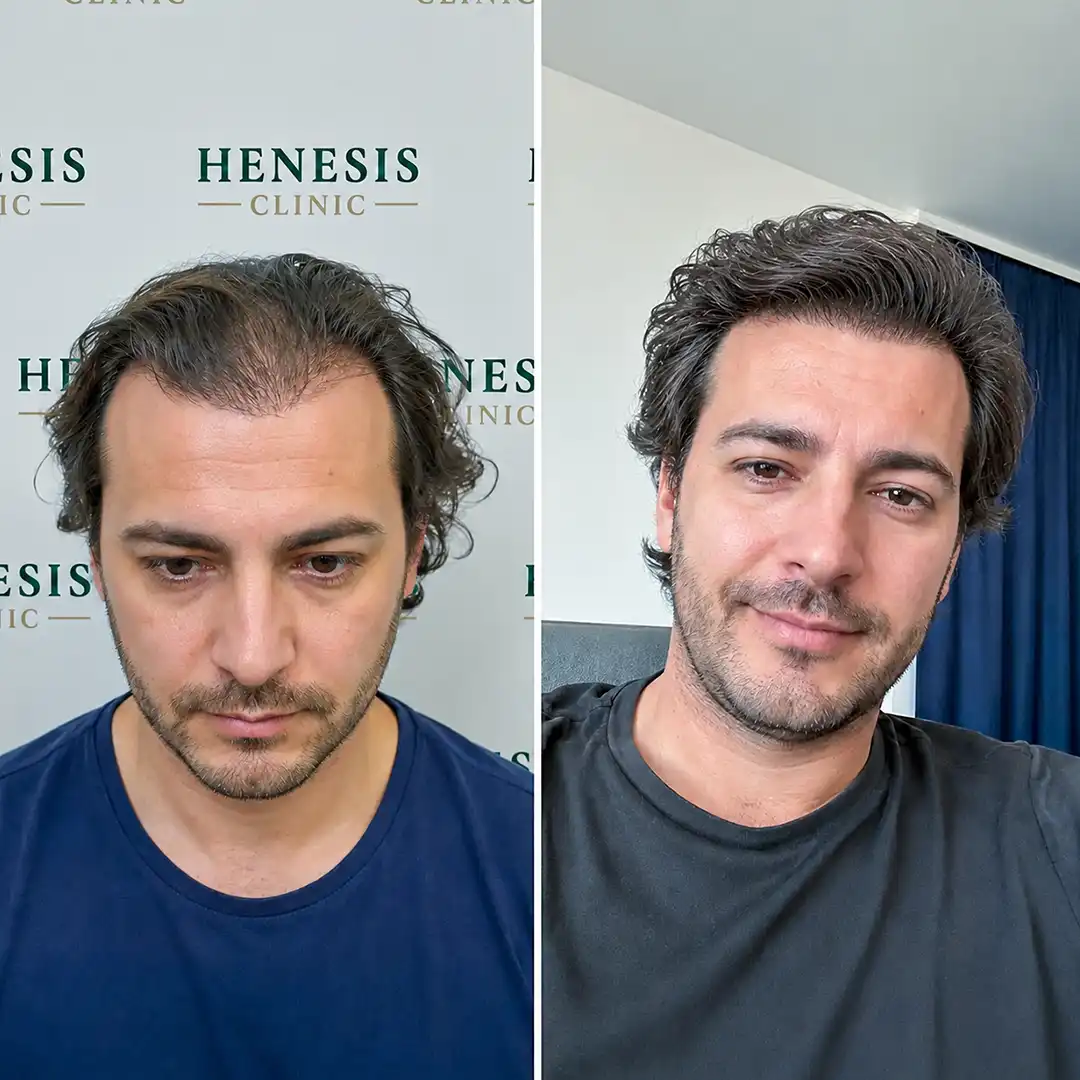
Before-treatment and 12-month follow-up photographs from documented case HNS-0073
Before Treatment 12-Month Follow-Up

Case record

Case at a Glance

Case ID
HNS-0073
Procedure
DHI Hair Transplant
Graft Count
2,700 grafts
Treatment Area
Frontal Hairline, Bitemporal Recessions, And Anterior Mid-Scalp
Hair Type
Caucasian / Dark Brown Wavy
Hair-Loss Pattern
Receding frontal hairline + with the patient maintaining longer existing hair
Donor Characteristics
Occipital donor area (concealed window-shaved extraction zone)
Procedure Date
28 August 2025
Follow-Up
12 months
Medical Doctor
Dr. Timur Aksu, MD
Clinic Location
Henesis Clinic · Istanbul, Türkiye

Documented case sequence

Case Timeline

  1. 01
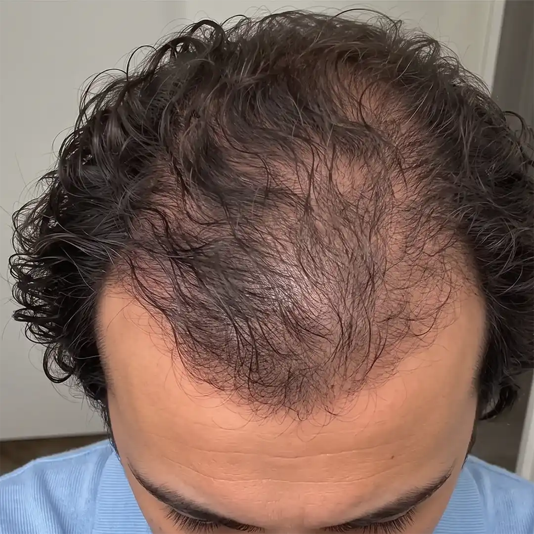
    Pre-treatment top view from documented case HNS-0073

    Pre-Treatment Assessment

    Frontal Hairline, Bitemporal Recessions, And Anterior Mid-Scalp planning

  2. 02
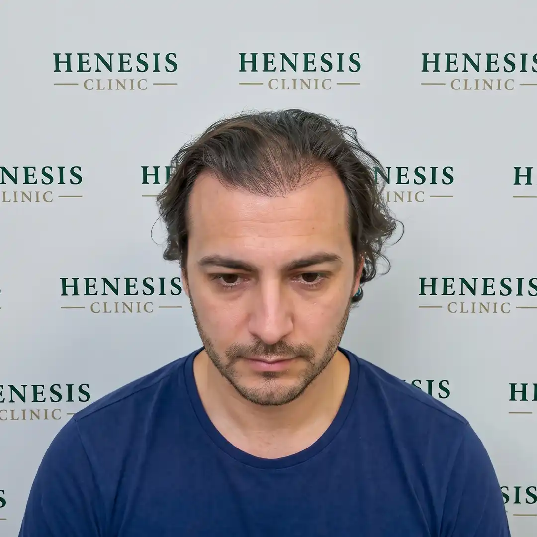
    Procedure front view from documented case HNS-0073

    Procedure

    28 August 2025

    DHI · 2,700 grafts

  3. 03
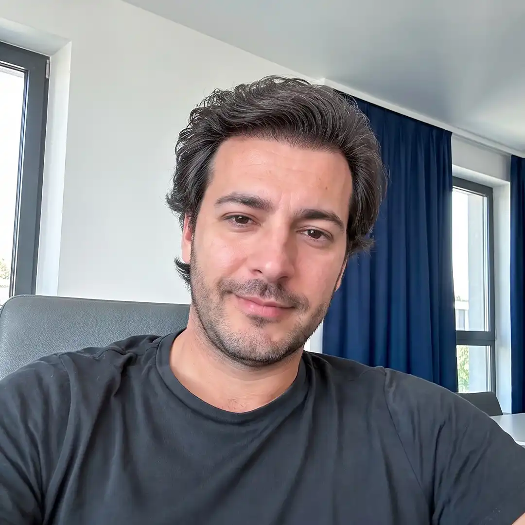
    12-month front-view follow-up from documented case HNS-0073

    12-Month Follow-Up

    Primary documented follow-up

    Follow-up photographs record the treated frontal hairline, temporal corners and mid-scalp.

  4. FINAL
    Before-treatment and 12-month follow-up photographs from documented case HNS-0073

    Before & After Comparison

    Baseline vs 12-Month Follow-Up

Documented clinical journey

Patient presentation and recorded decisions

Assessment & Treatment Planning

Patient goal: Lower and strengthen the frontal hairline, improve visible density in the temporal recession areas and increase forelock coverage while preserving the patient's longer hairstyle during treatment planning.

Assessment context: Receding frontal hairline, deep temporal recession and visible thinning across the central forelock documented in a Norwood 3V / 3A pattern, with the patient maintaining longer existing hair.

Planning rationale: An unshaven DHI approach was selected to allow graft implantation between existing longer native hairs without full shaving of the frontal recipient area. Donor harvesting was planned through a concealed window-shaved zone, while implantation strategy focused on angle, direction, depth and distribution according to the patient's native hair pattern.

Design notes: A structured masculine frontal hairline was planned according to the patient's facial proportions and treatment goals. Single-hair follicular units were prioritized along the leading edge to create a softer transition, with additional graft distribution planned through the temporal recession pockets and central forelock.

Recorded treatment details

Procedure Details

Technique
DHI
Donor Area
Occipital donor area (concealed window-shaved extraction zone)
Recipient Areas
Frontal Hairline, Bitemporal Recessions, And Anterior Mid-Scalp
Implantation Workflow
Implanter-assisted workflow

Recorded Procedure

Follicular units were harvested from a concealed window-shaved occipital donor zone using FUE-based micro-punch extraction. Selected grafts were loaded into Choi implanter pens and implanted across the frontal hairline, bitemporal recession zones and anterior mid-scalp with placement adapted for angle, direction, depth and distribution between existing native hairs.

Recorded follow-up context

Clinical Interpretation

Documented Observation

The pre-treatment scalp record describes receding frontal hairline, deep temporal recession and visible thinning across the central forelock documented in a Norwood 3V / 3A pattern, with the patient maintaining longer existing hair. The recorded treatment field was the frontal hairline, temporal corners and mid-scalp. The recorded procedure used 2,700 grafts. At 12 months, the clearest visual change is concentrated at the frontal hairline, with additional photographed change through the temporal corners and mid-scalp. The treatment did not target the crown, so these photographs should not be interpreted as evidence of crown restoration. An unshaven recipient strategy was recorded, so the images should be read in that context rather than as a fully shaved protocol. In the follow-up views, the transplanted areas appear visually continuous with the patient's existing wavy hair pattern. This is the documented result of one patient and does not predict another patient's outcome.

Follow-Up
12 months
Complications
None documented.
Further Treatment
No further treatment currently documented.

Medical doctor

Dr. Timur Aksu, MD

Dr. Timur Aksu, MD is the physician recorded for this case. View the physician profile for clinical focus, responsibilities and physician information.

View Doctor Profile

Your anatomy and goals are individual

Find Out What May Be Suitable for Your Hair

Share clear photos and information about your hair loss with the Henesis team for an individual assessment.

Request Assessment